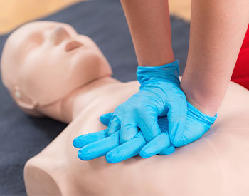
First Aid Training - Cardiopulmonary resuscitation. First aid course on cpr dummy.
Cardiopulmonary Services
The Cardiopulmonary Department at Mizell Memorial Hospital provides EKG, 24 and 48 hour Holter Monitor, and cardiac stress testing to patients. They also provide ultrasounds of the heart, carotid arteries, other vasculature. EKG's are done 7 days a week, 24 hours a day with an order form a physician. Other testing is scheduled Monday - Friday by appointment and only with an order from a physician.
Specific Procedures are listed below and further information can be read regarding your procedure:
*
Electrocardiogram (ECG)
*
Holter Monitor
*
Cardiac Stress Test
*
Echocardiogram (ECHO)
*
Nuclear Stress Test
*
Carotid Ultrasound
Electrocardiogram (ECG)
An electrocardiogram is used to monitor your heart. Each beat of your heart is triggered by an electrical impulse normally generated from special cells in the upper right chamber of your heart. An electrocardiogram - also called an ECG or EKG - records these electrical signals as they travel through your heart. Your doctor can use an electrocardiogram to look for patterns among these heartbeats and rhythms to diagnose various heart conditions. An electrocardiogram is a noninvasive, painless test. No special preparations are necessary.
During the test:
Ten electrodes will be attached to your arms, legs and chest. The electrodes are sticky patches applied with a gel to help detect and conduct the electrical currents of your heart. If you have hair on the parts of your body where the electrodes will be placed, the therapist may need to shave the hair so that the electrodes stick properly. You can breathe normally during the electrocardiogram. Moving, talking or shivering may distort the test results. A standard ECG takes just a few minutes.
The ECG is interpreted by a Cardiologist and the results are sent to your doctor.
Holter Monitor
A Holter monitor is a small, wearable device that records your heart rhythm. You usually wear a Holter monitor for 24 or 48 hours. During that time, the device will record all of your heartbeats. A Holter monitor test is usually performed after a traditional test to check your heart rhythm (electrocardiogram) isn't able to give your doctor enough information about your heart's condition. A Holter monitor has electrodes that are attached to your chest with adhesive and then are connected to a recording device. Your doctor uses information captured on the Holter monitor's recording device to figure out if you have a heart rhythm problem.
Wearing a Holter monitor may be a little inconvenient. The Holter monitor can't get wet, or it will be damaged. Don't swim or bathe for the entire time you're wearing your Holter monitor. While Holter monitors aren't usually affected by other electrical appliances, you shouldn't walk through a metal detector while wearing one. Holter monitoring is painless and noninvasive. You can hide the electrodes and wires under your clothes, and you can wear the recording device on your belt or attached to a strap. Once your monitoring begins, don't take the Holter monitor off - you must wear it at all times, even while you sleep.
Preparing for the test:
You should bathe before this appointment, because once your monitoring begins, you can't get the monitor wet or remove the monitor to bathe.
A therapist will place five to seven electrodes on your chest. If you have hair on the parts of your body where the electrodes will be placed, the therapist may need to shave the hair so that the electrodes stick properly.
The therapist will then connect the electrode to a recording device with several wires, so it can record data transmitted from the electrodes.
You'll be instructed to keep a diary of all the activities you do while wearing the monitor. It's particularly important to record in the diary any palpitations, skipped heartbeats, shortness of breath, chest pain or lightheadedness. Once your monitor is fitted and you've received instructions on how to wear it, you can resume your normal activities.
Once your monitoring period is over, you will return to the hospital and a therapist will remove the electrodes from your chest. This may cause some discomfort - similar to a bandage being pulled off your skin. You will turn in the diary you kept while you wore the Holter monitor. The Holter monitor is interpreted by a Cardiologist and the results are sent to your doctor.
Cardiac Stress Test
A cardiac stress test, also called an exercise stress test, is used to gather information about how well your heart works during physical activity. Because exercise makes your heart pump harder and faster than it does during most daily activities, an exercise stress test can reveal problems within your heart that might not be noticeable otherwise. A cardiac stress test involves walking on a treadmill while your heart rhythm, blood pressure and breathing are monitored.
Preparing for the test:
Wear comfortable clothes and good walking shoes to the exercise stress test. Before you start the test, a therapist places electrodes on your chest. If you have hair on the parts of your body where the electrodes will be placed, the therapist may need to shave the hair so that the electrodes stick properly. The electrodes are connected by wires to an electrocardiogram (ECG or EKG) machine. The electrocardiogram records the electrical signals that trigger your heartbeats. A blood pressure cuff is placed on your arm to check your blood pressure during the exercise stress test.
You then begin slowly walking on the treadmill and as the test progresses, the speed and elevation of the treadmill increases. A railing is provided on the treadmill that you can use for balance, but don't hang on to it, as that may cause artifact that can affect the results of the test.
The length of the test depends on your physical fitness and symptoms. The goal is to have your heart work hard for about eight to 12 minutes in order to thoroughly monitor its function. You continue exercising until your heart rate has reached a set target or until you develop symptoms that don't allow you to continue.
A typical exercise stress test lasts 15 minutes or less. You may stop the test at any time if you're too uncomfortable to continue exercising. After you stop exercising, you will be able to lie down with the monitors in place so that they can continue taking measurements as your heart rate and breathing return to normal. When your exercise stress test is complete, you may return to your normal activities for the remainder of the day. A Cardiologist will interpret your stress test and send the results to your doctor.
Echocardiogram (ECHO)
An echocardiogram uses sound waves to produce images of your heart which allows the Cardiologist to see how your heart is beating and pumping blood. The Cardiologists can use the images from an echocardiogram to identify various abnormalities in the heart muscle and valves. It gives the Cardiologist information about the size and shape of the heart along with estimates of the heart function such as cardiac output, ejection fraction and diastolic function.
Preparing for the test:
There are few risks involved in a standard echocardiogram. You may feel some discomfort similar to pulling off an adhesive bandage when the technician removes the electrodes placed on your chest during the procedure.
No special preparations are necessary for a standard echocardiogram.
After undressing from the waist up, you'll lie on a bed. The technician will attach electrodes to your chest to help detect and conduct the electrical currents of your heart.
During the echocardiogram, the technician may dim the lights to better view the image on the monitor. You may hear a pulsing "whoosh," which is the machine recording the blood flowing through your heart.
Most echocardiograms take less than 30 minutes, but the timing may vary depending on your condition. During an echocardiogram, you may be asked to breathe in a certain way or to roll onto your left side. Sometimes the transducer must be held very firmly against your chest. This can be uncomfortable - but it helps the technician produce the best images of your heart. A Cardiologist will interpret the echocardiogram and the results will be sent to your doctor.
Nuclear Stress Test
A nuclear stress test measures blood flow to your heart muscle both at rest and during stress on the heart. It's performed similarly to a cardiac stress test, but provides images that can show areas of low blood flow through the heart and areas of damaged heart muscle. A nuclear stress test usually involves taking two sets of images of your heart - one set during an exercise stress test while you're exercising on a treadmill or with medication that stresses your heart, and another set while you're at rest. A nuclear stress test is used to gather information about how well your heart works during physical activity and at rest.
Preparing for the test:
You will be asked not to eat, drink or smoke after midnight before a nuclear stress test. You can take your medications with a small sip of water, unless your doctor tells you not to.
Wear or bring comfortable clothes with you to the exercise stress test.
When you arrive for your nuclear stress test, you will be asked about your medical history.
A nuclear medicine called thallium or myoview is injected into your bloodstream through an intravenous (IV) line, usually in your hand or arm. After the injection you will need to wait 30 to 45 minutes for this substance to mix with your blood and travel to your heart. A special scanner similar to an X-ray machine - which detects the radioactive material in your heart - creates images of your heart muscle. Inadequate blood flow to any part of your heart will show up as a light spot on the images because not as much of the radioactive dye is getting there.
Next a therapist will place electrodes on your chest. If you have hair on the parts of your body where the electrodes will be placed, the therapist may need to shave the hair so that the electrodes stick properly. The electrodes are connected by wires to an electrocardiogram (ECG or EKG) machine. The electrocardiogram records the electrical signals that trigger your heartbeats. A blood pressure cuff is placed on your arm to check your blood pressure during the test.
If you're unable to exercise adequately, you may be injected with a medication that increases blood flow to your heart muscle - simulating exercise - for the test.
You then begin walking on the treadmill and as the test progresses, the speed and incline of the treadmill increases. A railing is provided on the treadmill that you can use for balance, but don't hang on to it tightly, as that may skew the results of the test. On a stationary bike, the resistance increases as the test progresses, making it harder to pedal.
The length of the test depends on your physical fitness and symptoms. The goal is to have your heart work hard for about eight to 12 minutes in order to thoroughly monitor its function. You continue exercising until your heart rate has reached a set target or you develop symptoms that don't allow you to continue. You may stop the test at any time if you are too uncomfortable to continue exercising.
After exercising, you will be injected again with the nuclear medicine and you will be encouraged to eat and drink something. After 30 to 45 minutes you will have a second set of images taken of your heart. This second set of images will let the Cardiologist compare the blood flow through your heart at rest and while you're exercising.
When your nuclear stress test is complete, you may return to your normal activities for the remainder of the day. A Cardiologist will interpret you test and the results will be sent to your doctor.
Carotid Ultrasound
A Carotid ultrasound is a safe, painless procedure that uses sound waves to examine the structure and function of the carotid arteries in your neck. Carotid arteries deliver blood from your heart to your brain and are located on each side of your neck. Carotid ultrasound is usually used to test for blocked or narrowed carotid arteries, which can indicate an increased risk of stroke.
A technician conducts the test with a small, hand-held device called a transducer. The transducer emits sound waves and records the echo as the waves bounce off tissues, organs and blood cells. A computer translates the echoed sound waves into a live-action image on a monitor. In a Doppler ultrasound, the information about the rate of blood flow is translated into a graph.
Preparing for the test:
Wear a comfortable shirt with no collar or an open collar.
Don't wear a necklace or dangling earrings.
You shouldn't need to make any other preparations.
You will be asked to lie on your back during the procedure. The ultrasound technician will adjust the position of your head to improve access to the side of your neck.
The ultrasound technician will apply a gel to your skin above the site of each carotid artery. The gel helps eliminate the formation of air pockets between your skin and the transducer. The technician then gently presses the transducer against the side of your neck in order for the instrument to send and receive sound waves.
You shouldn't feel any discomfort during the procedure. If you do, let the technician know.
A carotid ultrasound usually takes about 30 minutes. A Cardiologist will interpret the test and the results will be sent to your doctor.